Eukaryotic Cell 3d Model
The intricate world encapsulated within a single eukaryotic cell unfolds with breathtaking clarity in this meticulously crafted 3D representation. This model provides an unparalleled opportunity to explore the fundamental building blocks of complex life, offering a deep dive into the microscopic universe that powers every multicellular organism. Educators, students, and scientific communicators discover an invaluable resource for visualizing the complex interplay of cellular components, moving beyond static diagrams to a dynamic, explorable environment.
Eukaryotic Cell 3d Model – Unveiling the Cell’s Inner Sanctum
Delve into the core of biological complexity as you navigate the expertly rendered internal structures of this eukaryotic cell. Every organelle, from the expansive endoplasmic reticulum to the power-generating mitochondria, receives meticulous attention, ensuring anatomical accuracy and visual appeal. This detailed cross-section reveals how these specialized compartments collaborate, maintaining cellular function and supporting life processes.
The model’s design emphasizes clarity and pedagogical effectiveness, making complex biological concepts accessible. Users identify and understand the distinct roles each organelle performs, observing their spatial relationships within the cytoplasm. This visual precision transforms abstract textbook knowledge into a tangible, interactive learning experience.
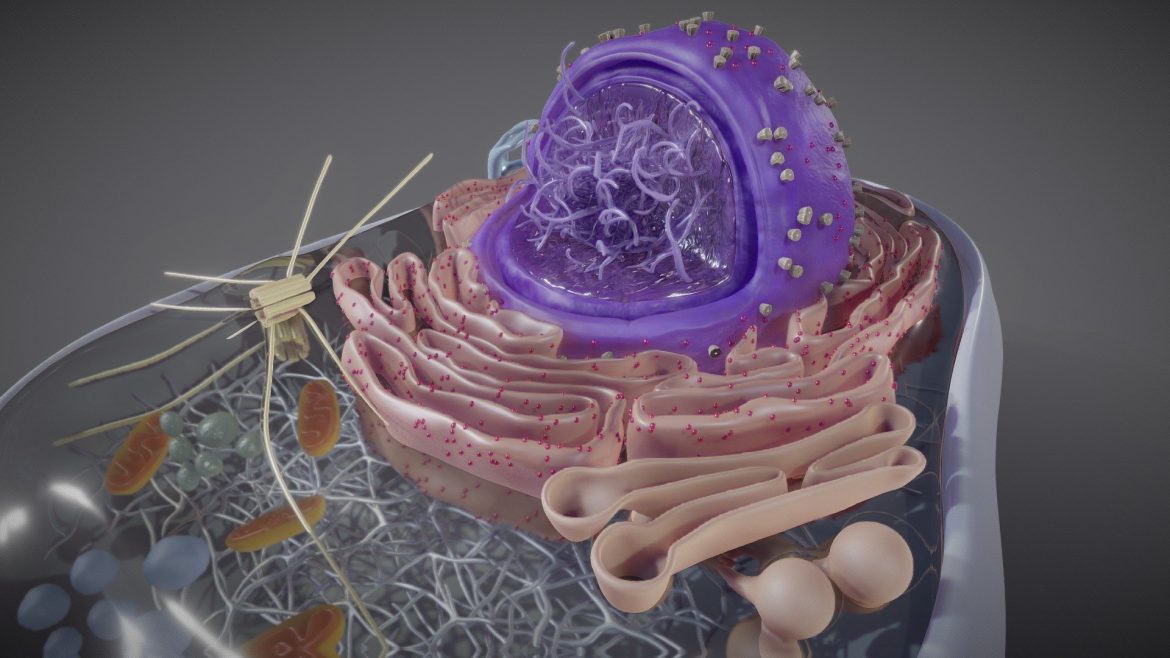
Eukaryotic Cell 3d Model – Precision in Microscopic Detail
Explore the subcellular landscape rendered with exceptional fidelity, highlighting features often difficult to discern in two-dimensional illustrations. The nucleus, with its intricate chromatin structure and prominent nucleolus, stands out as the cell’s command center. Ribosomes, vital for protein synthesis, appear precisely integrated, while lysosomes and peroxisomes demonstrate their crucial roles in waste management and detoxification.
This comprehensive visualization extends to the surrounding cellular environment, capturing the fluidity of the cell membrane and its selective permeability. Understanding these nuanced details becomes effortless, empowering users to grasp the sophisticated machinery driving cellular life. This truly offers an immersive glance into the microcosm.
Visualizing Life’s Fundamental Units
- Accurate representation of the nucleus, endoplasmic reticulum, Golgi apparatus, and mitochondria.
- Distinct visual differentiation of ribosomes, lysosomes, peroxisomes, and vacuoles.
- Clear depiction of the cell membrane and cytoplasm for contextual understanding.
- High-resolution textures and precise geometric modeling enhance educational impact.
This outstanding model serves as an indispensable tool for advanced biological studies, illustrating concepts from molecular biology to cellular physiology. Researchers use this resource to visualize hypotheses, while medical students gain a deeper appreciation for the cellular basis of health and disease. Its versatility supports various applications across scientific disciplines.
Elevating Biological Education
Integrate this exceptional visualization into your curriculum or research project to dramatically enhance engagement and comprehension. The high fidelity and scientific accuracy of this representation make it a benchmark for educational tools. It inspires curiosity and facilitates a profound understanding of the tiny, yet mighty, structures that constitute all living organisms.
For those seeking to explore life’s foundational components, this eukaryotic cell 3d model presents an unparalleled opportunity. It bridges the gap between theoretical knowledge and practical visualization, offering an intuitive platform for discovery. Discover more about cell biology on resources like Cell Press. For other high-detail models, consider checking out our Tango.
Seller Original Description
Animation showing the different componenets of an Eukaryotic Cell
Extended Use License
has been added to your cart!
have been added to your cart!
You must log in and be a buyer of this download to submit a review.
| ABOUT THE SELLER |
|
| SELLER-USER-NAME | Ebers |
| 3D Model formats | FBX, OBJ, BLENDER, TEXTURES, Materials |
| 3D Model details | VR / AR / Low-poly, Textures, Materials, UV Mapping, Scale transformations |
| Triangles | 1.2M |
| Vertices | 621.9k |
| Category | Science & Technology |
| Tags | anatomy, biology, cell, cross section, cytoplasm, ebers, education, eukaryotic cell, eukaryotic cell 3d model, human, medicine, micro, microbiology, microscopy, nature, nucleus, organelle, pharma, science |