Eukaryotic Cross Section 3d Model
Eukaryotic Cross Section 3d Model – Unveiling Microscopic Wonders
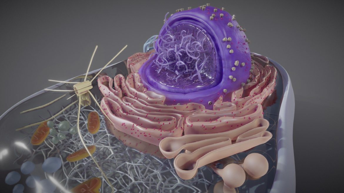
Explore the intricate internal landscape of a eukaryotic cell, meticulously rendered to highlight each vital component. This model serves as an indispensable visual aid, transforming abstract biological concepts into tangible, explorable forms. Witness the nucleus, the cell’s control center, alongside the energy-producing mitochondria and the protein-synthesizing ribosomes, all within their proper spatial relationships.
Every organelle, from the endoplasmic reticulum’s vast network to the Golgi apparatus’s processing hubs, stands out with remarkable definition. Users can visualize how these structures collaborate to sustain cellular life, offering a dynamic perspective often unattainable through traditional static diagrams. This depth of detail enhances comprehension and fosters a deeper appreciation for cellular biology.
Eukaryotic Cross Section 3d Model – Precision in Cellular Architecture
Experience unparalleled accuracy in this digital recreation, where expert 3D artistry meets scientific rigor. The model faithfully captures the nuanced morphology of a typical eukaryotic cell, ensuring that every membrane, pore, and internal structure aligns with current biological understanding. This commitment to precision makes it an authoritative resource for academic study and scientific illustration.
Students can confidently identify and differentiate between various organelles, solidifying their knowledge of cellular anatomy and function. Researchers benefit from a highly detailed visual reference, enabling them to communicate complex biological concepts more effectively. The model’s clarity supports deeper analytical engagement, making it a cornerstone for advanced biological education.
Key Features of the Model
This 3D model presents a comprehensive and visually rich exploration of cellular structure, designed for optimal learning and detailed examination. It provides a robust platform for understanding the intricate design of life at its most fundamental level.
- High-resolution textures render each organelle with distinct visual characteristics.
- Clearly delineated plasma membrane, cytoplasm, and nuclear envelope.
- Identifiable structures such as mitochondria, endoplasmic reticulum, and Golgi apparatus.
- Designed for intuitive exploration across various 3D viewing platforms.
Elevating Biological Understanding
Integrate this exceptional resource into your educational or professional toolkit to elevate biological understanding. Its detailed representation bridges the gap between theoretical knowledge and visual comprehension, making the complexities of cell biology accessible to a broader audience. The eukaryotic cell cross section 3d model serves as an indispensable resource for anyone seeking to master the fundamentals of life sciences.
This model stands as a testament to the power of digital visualization in scientific education, offering an engaging and effective way to learn about the building blocks of life. For more fascinating insights into the natural world, consider exploring resources like National Geographic Science. For other high-detail models, consider checking out our Hair – Male Spiky Anime model.
Seller Original Description
Animation showing the different componenets of an Eukaryotic Cell
Extended Use License
has been added to your cart!
have been added to your cart!
You must log in and be a buyer of this download to submit a review.
| ABOUT THE SELLER |
|
| SELLER-USER-NAME | Ebers |
| 3D Model formats | FBX, OBJ, BLENDER, TEXTURES, Materials |
| 3D Model details | VR / AR / Low-poly, Textures, Materials, UV Mapping, Scale transformations |
| Triangles | 1.2M |
| Vertices | 621.9k |
| Category | Science & Technology |
| Tags | anatomy, biology, cell, cytoplasm, ebers, education, eukaryotic cell, eukaryotic cell cross section 3d model, human, medicine, micro, microbiology, Mitochondria, nature, nucleus, organelle, pharma, science |